Celiac Disease Has a Little Sister – Dermatitis Herpetiformis
 Often sidelined as the skin manifestation of celiac disease, Dermatitis herpetiformis (DH) is the source of much physical – and emotional – aggravation for the estimated 15-25% of celiacs who struggle with it. Debilitatingly itchy and like celiac disease, often misdiagnosed, visible red bumps and raw blisters exact an additional emotional toll through stigmatization and avoidance of situations where skin can be seen by others.
What is DH? 1 
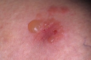
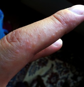
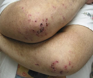
Dermatitis Herpetiformis bumps and blisters resemble herpes lesions, hence the name “herpetiformis”, but are NOT caused by the herpes virus. They typically appear on both sides of the body, most often on the forearms near the elbows, as well as on knees and buttocks. Symptoms of DH tend to come and go, and it is commonly diagnosed as eczema. Symptoms normally resolve when on a strict, gluten-free diet.
DH affects 15 to 25 percent of people with celiac disease who typically have no digestive symptoms. DH can affect people of all ages, but most often appears for the first time in those between the ages of 30 and 40. People of northern European descent are more likely than those of African or Asian heritage to develop DH. The condition is somewhat more common in men than women. And men are more likely to have atypical oral or genital lesions.
How does a disorder that damages the intestines show up on the skin?
 “When a person with celiac disease consumes gluten, the mucosal immune system in the intestine responds by producing a type of antibody called immunoglobulin A (IgA)”, explains John Zone, MD, CDF Medical Advisory Board Member and Chairman of the Department of Dermatology at the University of Utah School of Medicine. As IgA enters the bloodstream, it can collect in small blood vessels under the skin, triggering further immune reactions that result in the blistering rash of DH.
Diagnosing DH with Skin Biopsy and Blood Tests
A skin biopsy is used to confirm a diagnosis of DH. Dermatologists usually use what’s called a “punch biopsy” to remove the skin and test it for dermatitis herpetiformis. After injecting a local anesthetic, your dermatologist will use a tiny, cookie-cutter-like punch to remove a 4mm sample of skin. The incision can be closed with one stitch and generally heals with very little scarring.
A skin sample is taken from the area next to a lesion and a fluorescent dye is used to look for the presence of Immunoglobulin A (IgA) deposits that appear in a granular pattern. Skin biopsies of people with DH are almost always positive for this granular IgA pattern.
It is important to have your dermatitis herpetiformis skin biopsy performed by someone who has diagnosed the skin condition before and knows how to do the biopsy. The skin sample must be taken from skin directly adjacent to the suspected dermatitis herpetiformis lesion, as opposed to directly from the lesion, since inflammation in the lesion can destroy the IgA deposits.
Blood tests for other antibodies commonly found in people with celiac disease—antiendomysial and anti-tissue transglutaminase antibodies—supplement the diagnostic process. If the antibody tests are positive and the skin biopsy has the typical findings of DH, patients do not need an intestinal biopsy to confirm the diagnosis of celiac disease.
 Treatment for DH with Dapsone and the Gluten-Free Diet
If you are diagnosed with dermatitis herpetiformis, your dermatologist may prescribe dapsone for short-term relief from the itching. According to Dr. Zone, the rash responds dramatically to dapsone, usually in 48 to 72 hours. People who can’t tolerate dapsone may be given sulfapyridine or sulfamethoxypyridazine instead, although these drugs are less effective.
However, you’ll still need to follow a strict gluten-free diet to control your dermatitis herpetiformis. Even with a gluten-free diet, dapsone or sulfapyridine therapy may need to be continued for 1–2 years to prevent further DH outbreaks. In some cases, a diet high in iodine may worsen DH symptoms. If you are experiencing DH flareups, you should consult with a dermatologist expert in celiac disease, to determine if foods or medicines high in iodine are the cause.
Learn more about the Gluten-Free Diet from the Celiac Disease Foundation
From the Canadian Celiac Association:
Dermatitis herpetiformis (DH) is a chronic skin condition with a characteristic pattern of lesions, with intense itching and burning sensations.
Causes: Genetic factors, the immune system, and a sensitivity to gluten play a role in this disorder. The precise details remain unknown.
Incidence: DH is not uncommon. It affects males and females equally and occurs in about 1:100,000 people. It is more common in Caucasians than Blacks, and rare in the Japanese population. Onset is most frequently in the late second to the fourth decades of life.
Characteristics: A new unscratched lesion is red, raised, and usually less than 1 cm in diameter with a tiny blister at the centre. However, if scratched, crusting appears on the surface. The ‘burning” or “stinging” sensation is different from a “regular” itch, and can often occur 8-12 hours before a lesion appears.
Areas Affected: The most common areas are the elbows, knees, back of the neck, scalp, the upper back, and the buttocks. Facial and hair-line lesions are not uncommon; the inside of the mouth is rarely affected. The rash has symmetric distribution.
Diagnosis: Dermatitis herpetiformis is only diagnosed and confirmed by a dermatologist obtaining a slight skin biopsy from uninvolved skin adjacent to blisters or erosions. Other forms of dermatitis can mimic dermatitis herpetiformis necessitating skin biopsy for correct diagnosis.
Small bowel biopsies will confirm a diagnosis of coexisting celiac disease but are not essential if the skin biopsy confirms the diagnosis of dermatitis herpetiformis. Referral to a gastroenterologist may be necessary for assessing the extent of the underlying intestinal injury and associated deficiencies of iron, calcium and vitamins.
The skin symptoms usually predominate over intestinal symptoms. Blood tests for celiac disease may be negative, reflecting the absence or paucity of intestinal symptoms expected when there is milder, more patchy villous atrophy seen on small bowel biopsies.
Management: Treatment is by drugs, and diet restrictions.
• Drugs: Dapsone (Avlosulfon) or related sulphones: The response is dramatic. Within 24-48 hours the burning is relieved and the rash starts to disappear. The aim is to use the smallest dose possible to keep the itch and rash under control. It has no effect on the gut abnormality.
• Diet: Gluten-free Diet: Elimination of all wheat, rye, barley, oats, triticale, and any parts thereof from the diet will result in:
- the skin lesions improving
- the gut returning to normal
- a substantial reduction in or the elimination of the need for sulphones to control the skin rash
- a decreased risk of malignancy


